Describe the External and Internal Structure of a Kidney
In the concave side there is a depression in the middle point which is called as hilum or hilus. Tubule reabsorb the useful material like glucose amino acid salts and water into blood capillaries.

Kidney Structure Biology For Majors Ii
Renal cortex- outer granular shell.
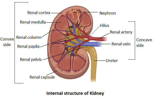
. Upper portions of the kidneys are somewhat protected by the eleventh and twelfth ribs Figure 1. The bean-shaped kidneys have an outer convex side and an inner concave side called the renal hilus where the renal artery vein and ureter are found. Each kidney weighs about 125175 g in males and 115155 g in females.
The kidneys are two bean-shaped organs found in vertebrates. The kidneys are bean-shaped with the convex side of each organ located laterally and the concave side medial. Describe the external and internal structure of a kidney.
Externally the kidneys are surrounded by three layers illustrated in Figure 2. Describe the anatomical structure of the ureterand its location in the body. A light outer area called the renal cortex and a darker inner area called the renal medulla.
The renal fat pad is a shock absorbing layer of adipose tissueThe renal capsule is the blood filtering region of the nephron located in the. The left kidney is located at about the T12 to L3 vertebrae whereas the right is lower due to slight displacement by the liver. Upper portions of the kidneys are somewhat protected by the eleventh and twelfth ribs Figure 257.
The outermost layer is a tough connective tissue layer called the renal fascia. Uretha- urine from bladder to outside of body. Renal medulla- inner mass of tissues.
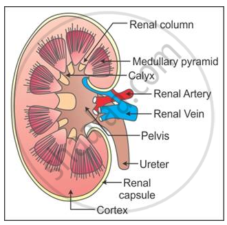
The kidneys are bilateral organs placed retroperitoneally in the upper left and right abdominal quadrants and are part of the urinary system. The cortex which extends between the medullary pyramids is called columns of Bertini. Internally it is divided into an outer cortex and an inner medulla.
A longitudinal section of the kidney shows two main regions-an outer dark cortex and an inner lighter medullaThe medulla is composed of finely striped substance arranged in several pyramidsThe. The renal artery renal vein and ureter enter the kidney via the hilus. Medullary pyramids are projected into the calyces.
The second layer is called the perirenal fat capsule which helps anchor the kidneys in place. Describe the external and internal anatomical structure of the kidney. Renal capsule- in renal cortex and medulla.
The left kidney is located at about the T12 to L3 vertebrae whereas the right is lower due to slight displacement by the liver. Each kidney weighs about 125175 g in males and 115155 g in females. The kidney is a bean-shaped structure.
Each kidney weighs about 125175 g in males and 115155 g in females. The outer edge of the kidney is convex and the inner concave. Upper portions of the kidneys are somewhat protected by the eleventh and twelfth ribs.
Renal External Anatomy Kidney. The kidneys have been bean-shaped appearance. Externally the kidney is surrounded by three layers which are the renal fascia the perirenal fat capsule and the renal capsuleThe fascia serve to firmly anchor the kidney to the posterior abdominal wall in a retroporitoneal position.
Internal structure - at first dirty blood enters into renal artery after that it reaches to glomerulus. The left kidney is located at about the T12 to L3 vertebrae whereas the right is lower due to slight displacement by the liver. Answer to Solved Describe the external structure of the.
This capsule maintains the kidneys shape and protects the inner tissues. In the medulla 5-8 renal pyramids are separated by connective tissue renal columns. A thin connective tissue called the renal capsule surrounds each kidney.
Describe the structure of the urinary bladder and its relations to the peritoneum. The internal structure of the kidney is divided into two main areas. Kidney Anatomy External.
I have some kidney issues myself--PKD type 3 or 4--one of the ones we dont yet have traced to a specific chromosome recurring stones and an acid-potassium imbalance. Dave Ward 16y. Describe the external structure of the kidney including its location support structures and covering Identify the major internal divisions and structures of the kidney Identify the major blood vessels associated with the kidney and trace the.
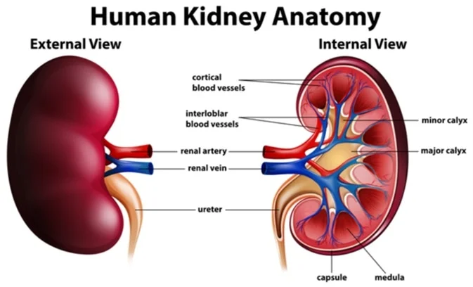
I just started a flickr group for people with any kind of kidney problems. Outer convex side and the inner concave side called the renal hilus this provides space for the renal artery vein and ureter to get to the kidney. The kidneys are paired bean-shaped organs that are located behind the abdominal peritoneum along with the posterior body wall.
Their shape resembles a bean where we can describe the superior and inferior poles as well as the major convexity pointed laterally and the minor concavity pointed medially. The medulla is in the form of medullary pyramids. After that remains enters into Bowmans capsule and after it reaches tubule.
Here the blood vessels of the kidney ie. Discuss the anatomical structure of urethraand the. A frontal section through the kidney reveals an outer region called the renal cortex and an inner region called the renal medulla Figure 2512.
Renal artery enters and Renal vein comes out of the kidney. External- reddish brown bean shaped smooth surface. Editor Aug 30 2017.
They are located on the left and right in the retroperitoneal space and in adult humans are about 12 centimetres 47 in in length. Covering the surface of each kidney is a tough semi-transparent connective tissue membrane. Glomerulus filters the dirty bloodsAnd blood cells also stop here.
The kidney and its vessels are embedded in a mass of fatty tissue called the perirenal fat which extends into a central cavity the renal sinus. Due to the size of the liver the right kidney is somewhat lower than the left kidney. Within the medulla there are eight 8 or more cone-shaped sections known as renal pyramids.
A thin layer of fibrous connective tissue forms the renal capsule surrounding the kidney. Please refer to diagram on right.

25 1 Internal And External Anatomy Of The Kidney Anatomy Physiology
Explain The Internal Structure Of The Kidney Class 11 Biology Cbse
Anatomy Of The Kidney
What Is The Internal Anatomy Of A Kidney Quora

25 1 Internal And External Anatomy Of The Kidney Anatomy Physiology

External And Internal Structure Of Kidneys Definition Examples Diagrams
Explain Internal Structure Of Kidney With The Help Of Suitable Diagram Biology Shaalaa Com

25 1 Internal And External Anatomy Of The Kidney Anatomy Physiology
What Is The Internal Anatomy Of A Kidney Quora


Comments
Post a Comment